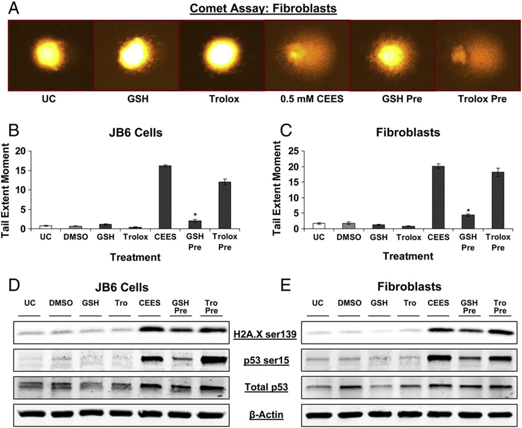
Fig. 6.
Effects of GSH or Trolox treatment on CEES-induced DNA damage in JB6 cells and fibroblasts. (A–C) JB6 cells and fibroblasts were treated with 10 mM GSH or 800 µM Trolox for 1 h before 0.5 mM CEES exposure for 1 h; cells were thereafter collected and subjected to comet assay as described under Materials and methods. (A) Representative pictures of the comet showing the effects of the GSH or Trolox treatment in fibroblasts. (D and E) JB6 cells and fibroblasts were treated with 10 mM GSH or 800 µM Trolox for 1 h before 0.5 mM CEES exposure for 4 h; whole-cell lysates were prepared at the end of the treatments and subjected to SDS–PAGE followed by Western blot analysis as described under Materials and methods to detect H2A.X Ser139 and p53 Ser15 phosphorylation. Membranes were stripped and reprobed with total p53 and β-actin antibodies. Data are presented as means ± SEM, n = 3. *P<0.001 compared to 0.5 mM CEES treatment. UC, untreated control; DMSO, vehicle control.

