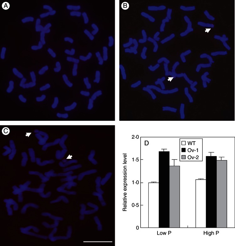
Fig. 4.
Fluorescent in situ hybridization (FISH) and expression analysis of Ta-PHR1-A1 in the wild type (WT) and its transgenic lines. (A–C) pUBI::Ta-PHR1-A1 vector was used as the detection probe in FISH of the wild type (A) and the transgenic lines Ov-1 (B) and Ov-2 (C). Fluorescent signals are indicated by arrows. Scale bar = 10 µm. (D) Relative expression levels of Ta-PHR1 in the WT and the transgenic lines Ov-1 and Ov-2 grown in nutrient solution. Seeds were germinated for 7 d, and then the germinated seeds with residual endosperm removed were transferred to nutrient solution containing 10 µm Pi (low P) and 200 µm Pi (high P) for 7 d, respectively. Then the seedlings were collected for gene expression analysis. Data are the mean ± s.e. of three biological replications.

