
Figure 1.
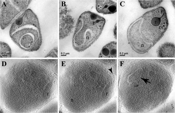
Structural organization of merozoites processed by different electron microscopy techniques. A to C – different examples of merozoites processed by conventional transmission electron microscopy. Rhoptries (r), micronemes (m) and the nucleus (n) are clearly observed. D to F – different sections from a tomogram of a cryo-preserved merozoite. Although more difficult to identify, some structures stand out like the rhoptries (r), the polar rings (arrowhead) and the apicoplast (arrow).