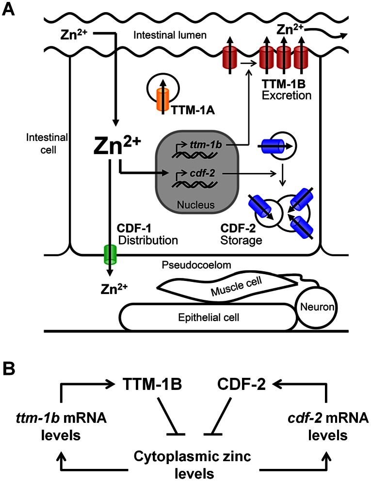
Figure 9. Network of CDF zinc transporters in intestinal cells.
(A) Dietary zinc moves from the intestinal lumen to the cytoplasm of intestinal cells by an undefined mechanism. High levels of cytoplasmic zinc increase transcription of ttm-1b and cdf-2. CDF-1 (green) functions in distribution; it is localized to the basolateral surface of the plasma membrane of intestinal cells and transports cytoplasmic zinc into the pseudocoelum for use by cells such as epithelia, muscles and neurons. TTM-1B (red) functions in excretion; it is localized to the apical surface of the plasma membrane of intestinal cells and transports cytoplasmic zinc into the intestinal lumen. CDF-2 (blue) functions in storage: it is localized to the membrane of gut granules, lysosome-related organelles that acquire a bilobed morphology in response to high zinc, and it transports zinc into the lumen of these organelles [30]. TTM-1A (yellow) is localized to vesicles and may also promote zinc excretion and/or sequestration. (B) A parallel negative feedback circuit promotes zinc homeostasis in intestinal cells. TTM-1B and CDF-2 proteins reduce the level of cytoplasmic zinc and promote zinc detoxification by distinct mechanisms of zinc excretion and zinc sequestration, respectively. ttm-1b and cdf-2 transcripts are both induced by high levels of cytoplasmic zinc. Mutations that reduce the activity of ttm-1 or cdf-2 cause induction of the remaining protein.

