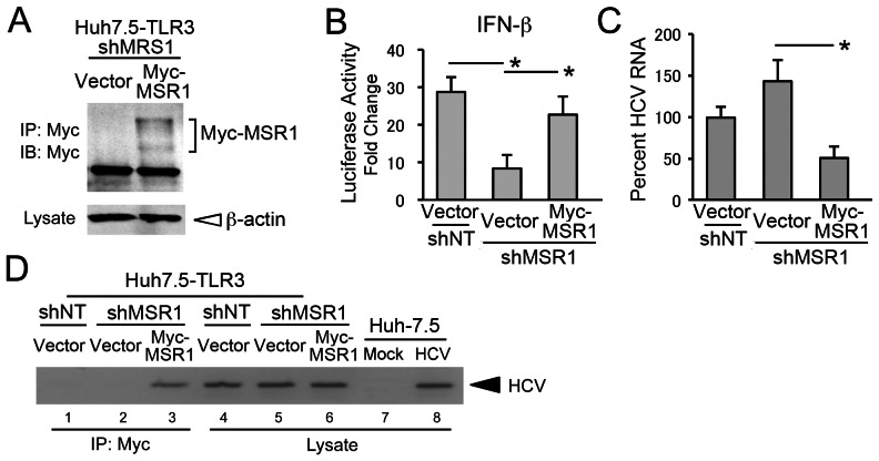
Figure 4. Reconstitution of MSR1 expression in MSR1-depleted cells restores TLR3 signaling triggered by HCV.
(A) Reconstitution of MSR1 expression in Huh7.5-TLR3/shMSR1 cells. Myc-tagged MSR1 was stably expressed by retroviral transfer of a shMSR1-resistant vector (pCX4bsr/Myc-MSR1). Lysates were immunoprecipitated with anti-Myc, then subjected to immunoblotting as described in Materials and Methods. “Vector” = cells transduced with empty vector. (B) Restoration of poly(I:C) induction of IFN-β promoter activity by ectopically expressed Myc-MSR1 in Huh7.5-TLR3/shMSR1 cells. Cells were co-transfected with pCX4bsr/Myc-MSR1 (vs. empty vector), pIFN-β-Luc and pRL-CMV (internal control reporter) and cultured for 24 h, then treated with poly(I:C) (50 µg/ml) for 6 hrs prior to lysis and luciferase assay. Huh7.5-TLR3/shNT cells, transfected with empty vector, were included as a positive control. (C) Quantitative RT-PCR analysis of HCV RNA in Huh7.5-TLR3/shMSR1 cells stably expressing Myc-MSR1 (vs. empty vector) 72 hrs after infection with HJ3-5 virus at an m.o.i. of 1. (D) Co-immunoprecipitation of HCV RNA with Myc-MSR1 in lysates of Huh7.5-TLR3/shMSR1 cells stably expressing Myc-MSR1 (lanes 3 and 6) vs. empty vector (lanes 2 and 5). Cells were infected with HJ3-5 virus (m.o.i. = 1) 72 hrs prior to lysis. Immunoprecipitation was with anti-Myc, demonstrating association of HCV RNA with Myc-MSR1. See legend to Fig. 1E for further details.

