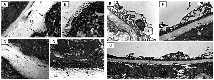
Figure 3. TEM of metacestodes recovered from untreated and subcutaneously infected treated mice.
Metacestodes recovered from intraperitoneally (A and B) and subcutaneously (C and D) infected mice exhibit a well-defined laminated layer (LL), which is in close physical contact with adjacent host tissue (ht). The tegument, with microtriches (arrows) protruding well into the LL, and the germinal layer (GL) exhibiting undifferentiated cells and glycogen storage cells, are clearly visible. Metacestodes from animals subcutaneously infected and treated orally with albendazole show a reduced and less defined laminated layer (LL), which maintains contact with host tissue components. This is clearly visible in G. Other alterations can be seen in E and F, such as the loss of integrity of the germinal layer (GL) and the absence of microtriches (for comparison refer to A–D). Similar structural damage was observed on the material collected from intraperitoneally infected animals treated with albendazole (results not shown).

