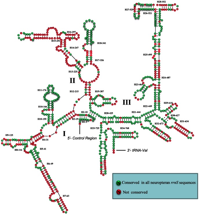
Figure 3. Secondary structure of T. langii rrnS.
Each helix is numbered progressively from the 5′ to 3′ end together with the first nucleotide belonging the helix itself. Domains are labeled with Roman numbers. Dashes (−) indicate Watson-Crick base pairing and dots (•) indicate unmatched base pairing.

