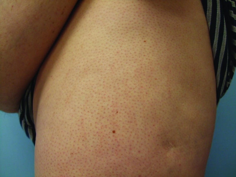
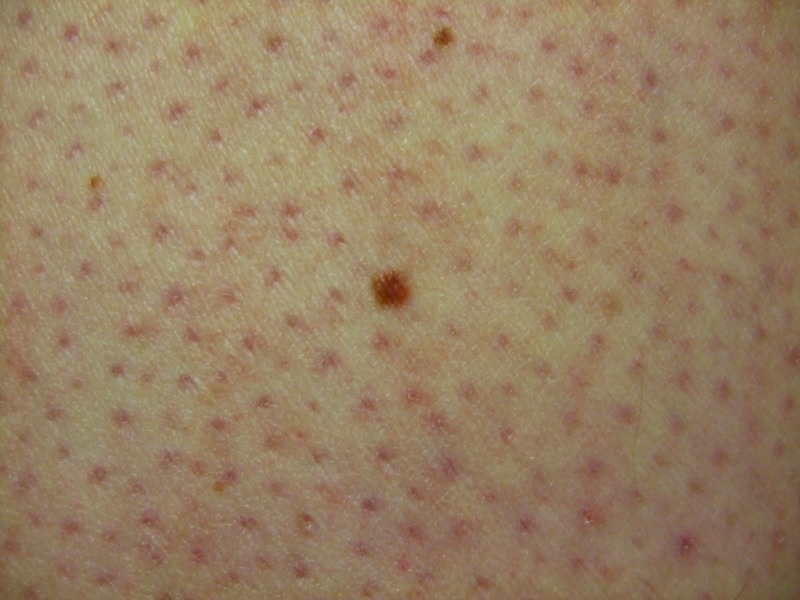
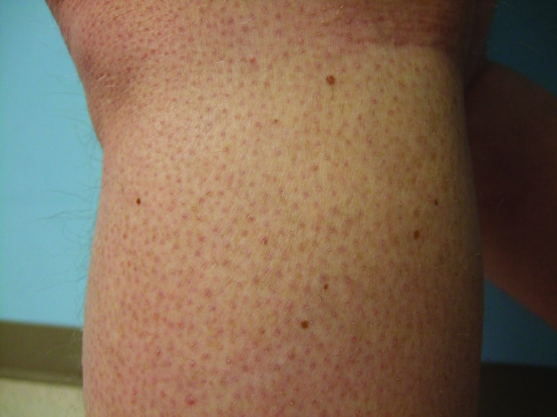
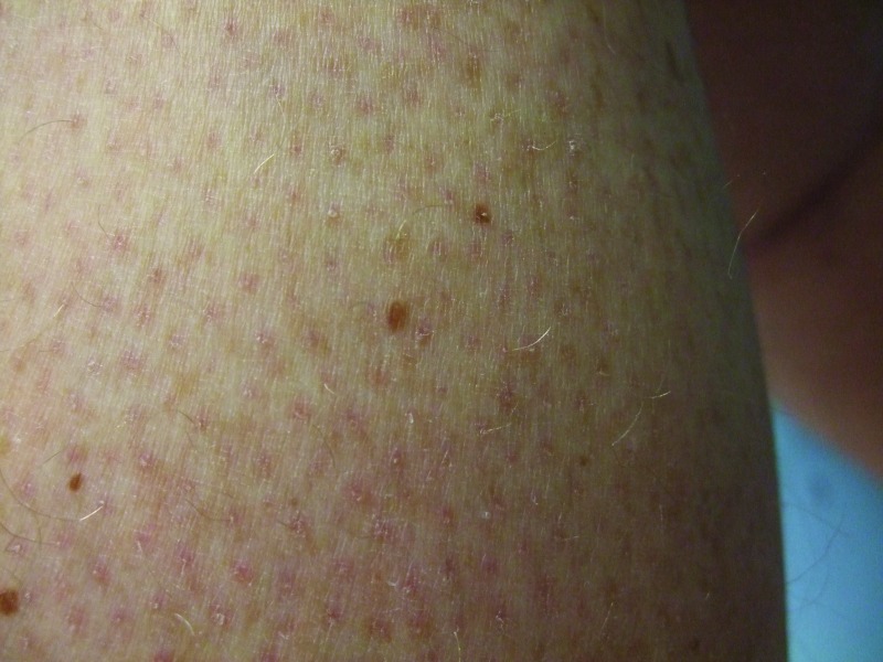
Figures 5A, 5B, 5C, and 5D.
Distant (A and C) and closer (B and D) views of the left lateral thigh (A and B) and left calf (C and D) show the vemurafenib-associated new melanocytic nevi that were biopsied; these were the inferior and larger brown lesion on the thigh (B) and the superior and lateral brown lesion on the calf. Pathology analysis of each pigmented lesions showed a dysplastic nevus characterized by a compound melanocytic nevus with moderate architectural disorder and moderate cytologic atypia. His keratosis pilaris-like eruption is also prominent.


