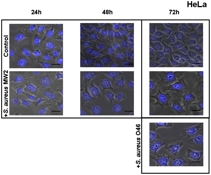
Figure 2. Enlargement of HeLa cells exposed to S. aureus.
Human HeLa cells were exposed for 2 h to S. aureus MW2 or O46 at MOI 20∶1 and further incubated for 24 h, 48 h and 72 h. After incubation, the cells were fixed, stained with DAPI and observed using ×400 magnification. The merged image of phase contrast and DAPI-stained cells is presented. Red arrows indicate the enlarged cells in infected cell cultures. Scale bars: 10 µm. A. Cells exposed to S. aureus MW2 for 24 h, 48 h and 72 h at MOI 20∶1. B. Cells exposed to S. aureus O46 for 72 h at MOI 20∶1.

