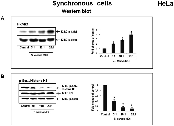
Figure 7. S. aureus-induced accumulation of phosphorylated Cdk1 and unphosphorylated Ser10 Histone H3.
HeLa cells were synchronized by DTB and were exposed to S. aureus bacteria (MW2) at MOIs ranging from 5∶1 to 20∶1 for 2 h, followed by incubation in cDMEM-Gent100 for 2 h, and further incubated for 20 h. Cells were then suspended in Laemmli loading buffer, and Western blot analysis, either with anti-phospho-Cdk1 antibodies or anti-p-Ser10 Histone H3 antibody, was performed as described in the Materials and Methods section. The chemiluminescence reaction was visualized and processed with a G:BOX imaging system. Blots are representative of three separate experiments. Data are presented as mean ± SD from three densitometry scans. Tukey's Honestly Significant Difference test was applied for comparison of means between the groups. (*) P-values <0.05 compared with the control were considered to be significant.

