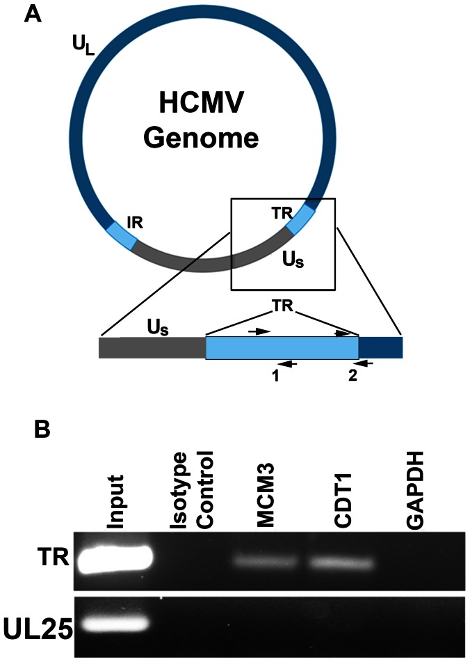
Figure 9. Interaction of MCM3 and CDT1 with HCMV TR DNA sequences in latently infected CD14 (+) monocytes.
Latently infected cells were treated with formaldehyde and subjected to ChIP using antibodies specific for MCM3 or CDT1. (A) Schematic of the HCMV genome showing the terminal repeat (TR) region. Also shown are primer sets specific for the TR segment of the genome (1 and 2). (B) ChIP assay showing an interaction of the TR region of the genome with MCM3 and CDT1 in latently infected cells. Control immunoprecipitations were the use of an isotype antibody control and an antibody specific for GAPDH. Control PCR primers were specific for the HCMV UL25 ORF.

