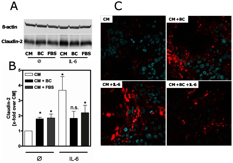
Figure 2. Changes of claudin-2 protein expression in intestinal epithelial cells.
(A) Western blot analysis revealed elevated claudin-2 expression in 14 days postconfluent differentiated Caco-2 cells after 48 h incubation with bovine colostrum (BC) or fetal bovine serum (FBS) compared to complete media (CM). β-actin was used as internal control for equal protein loading. (B) Relative expression of claudin-2 protein in Caco-2 cells was increased upon 48 h pre-incubation with BC compared to CM and after stimulation with IL-6 (50 ng/ml) for 24 h. *p<0.05 vs. CM, n = 5–6. (C) Immunofluorescence of Caco-2 cells after incubation with BC confirmed elevation of claudin-2 (red) by BC and IL-6 (24 h, 50 ng/ml) compared to CM after 48 h. Nuclei were counterstained with 4′,6-diamidino-2-phenylindole (blue). Representative images of 3 individual experiments. Magnification: ×600.

