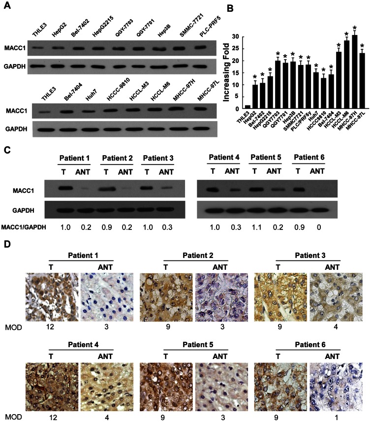
Figure 1. Expression of MACC1 is elevated in HCC.
(A-B) protein (A) and mRNA (B) expression of MACC1 in THLE3 and 15 cultured liver cancer cell lines. GAPDH was used as a loading control. (C-D) Western blot (C) and IHC (D) analysis of MACC1 protein in each of the primary liver cancer tissue (T) and adjacent non-cancerous tissues (ANT) samples taken from the same patient. Error bars represent mean ± SD from three independent experiments. * P<0.05.

