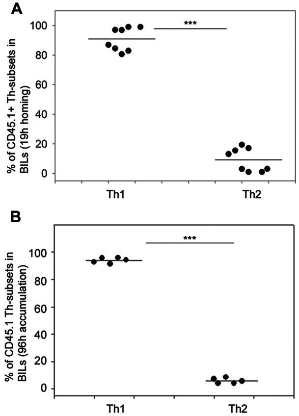
Figure 2. Adoptively transferred Th1 cells show preferential homing and accumulation in an intracranial tumour compared with Th2 cells.

SMARTA T cells (CD45.1) were labelled with CFSE (Th1) or Violet dye (Th2) and were intravenously transferred (3×10⁶ Th1; 3×10⁶ Th2) into C57BL/6 mice (CD45.2) that had been intracranially implanted with 4×10⁵ MC57-GP tumour cells 4 days previously. After 19 hours (A), or 96 hours (B), BILs were isolated, stained with antibodies for CD4 and CD45.1 and were analysed ex vivo by multicolour flow cytometry. Adoptively transferred T cells were identified as CD45.1+CD4+ cells that were either CFSE+ or Violet dye+ (supporting information Figure S2). Results are expressed as the percentage of Th1 and Th2 cells among the adoptively transferred CD45.1+CD4+ cells in the BILs, each symbol represents an individual mouse (n = 8). ***P<0.001, t-test.
