
Figure 5. CD40L expressing mCD8+ T cells promote secondary expansion following homologous boost.
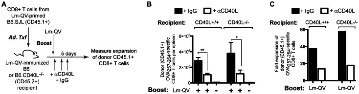
(A) Experimental design: Donor (B6.SJL) mice and recipient (C57BL/6 and CD40L-deficient) mice were concurrently primed with 107 cfu Lm-QV. 21 days later, CD8+ T cells were purified from B6.SJL spleens and transferred into B6 and B6.Cd40L-/- mice. 24 h post-transfer, recipients were boosted with 5×10⁶ cfu Lm-QV in the presence of αCD40L or control antibody. OVA257–264-specific memory CD8+ T cell responses were assessed 5 days following boost using intracellular cytokine staining for IFN-γ (n = 5). (B) Total OVA257–264-specific (IFN-γ+) CD45.1+CD8+ T cells per animal (mean, ±SEM). *, P<0.05, **, P<0.01, Mann-Whitney. (C) Fold expansion of donor CD45.1+ IFN-γ + OVA257–264-specific CD8+ T cells. Data are representative of three independent experiments.