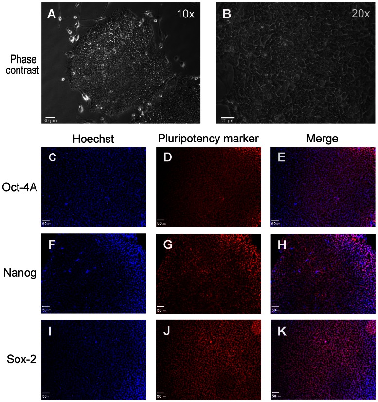
Figure 1. In vitro culture of vector-free hiPS cells in mTeSR1.
(A, B) Vector-free hiPS cells cultured in mTeSR1 media on human ES qualified matrigel under serum- and feeder-free conditions, show a typical pluripotent cell morphology growing in colonies as monolayers. (B) is a magnified image (20x) of (A). (C to K) hiPS cells express pluripotency markers Oct-4A (C, D and E), Nanog (F, G and H) and Sox-2 (I, J and K). Nuclei were labeled with the nucleic acid counter-stain Hoechst-33342. Bar = 30 µm for A, 20 µm for B and 50 µm for C to K.

