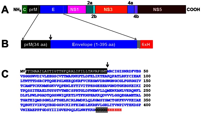
Figure 1. Design of the DENV-2 E antigen.
(A) Schematic representation of the DENV-2 polyprotein, showing the parts of prM and E included in designing the E antigen for expression in P. pastoris. (B) Design of the DENV-2 E antigen consisting of the 395 aa residue E ectodomain, preceded by the C-terminal 34 aa residues of prM. The grey box denotes the pentaglycine linker peptide joining the C-terminus of E ectodomain to the polyhistidine tag (6×H). (C) The predicted aa sequence of the DENV-2 E antigen shown in ‘B’. The color scheme corresponds to that shown in ‘B’. The first two aa residues (MV) were introduced due to the insertion of the initiator codon in a Kozak consensus context. The downward arrows in ‘B’ and ‘C’ denote the signal cleavage site.

