
Figure 4. Electron microscopic analysis of purified DENV-2 E protein.
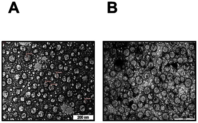
(A) Freshly purified DENV-2 E antigen was negatively-stained with uranyl acetate and examined under EM. (B) EM analysis was carried out after incubating the purified antigen at 37°C for 2 weeks.