Figure 2.

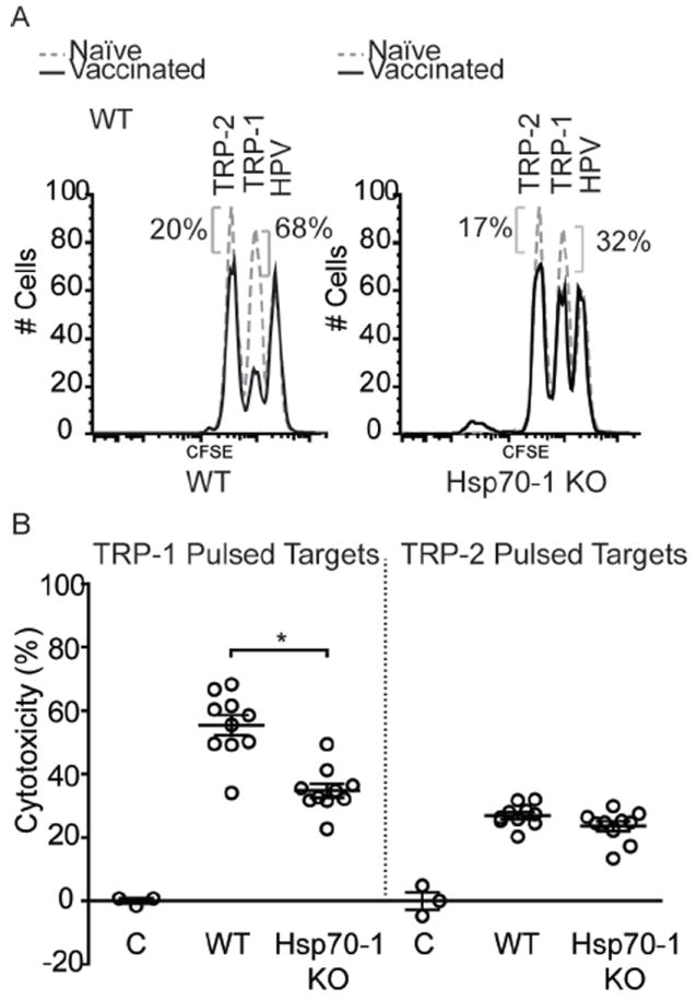
CTL killing towards melanoctye antigens is reduced in Hsp70-1 knockout mice. (A) For in vivo cytotoxicity assays, mice from Figure 1 were challenged with splenocytes pulsed with immunodominant peptides from TRP-1 or TRP-2, or irrelevant control peptides plus differing concentrations of CFSE. Spleens were harvested 18 hours after for analysis of CTL activity by FACS. Data from individual wild-type (WT) and Hsp70-1 KO mice are shown. (B) 20.6% more cytotoxicity was observed in wild-type mice (55.4%) towards TRP-1 pulsed splenocytes than in Hsp70-1 KO mice (34.8%). Approximately 25% cytotoxicity towards TRP-2-pulsed splenocytes is indicative of epitope spreading. Peptide pulsed splenocytes from control naïve mice are depicted as (C). Data are presented as means ± SEM. (*P < 0.05; n = 10 per group.)
