Figure 3.

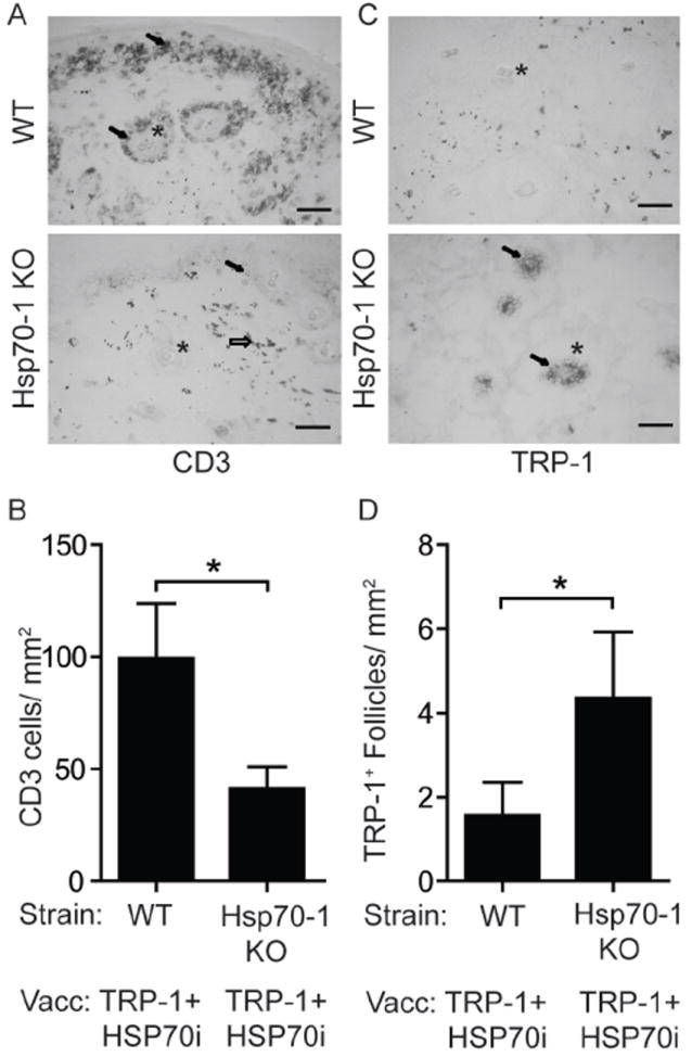
Immunohistology indicates inducible HSP70 is necessary for T cell-mediated loss of melanocytes. Treatment of mice engaged in this experiment is described under Figure 1. (A) Image of skin near the dermo-epidermal junction from mice one week after the final booster gene gun vaccination. CD3+ T cells (arrows) are more abundant near hair follicles (*) of wild-type (WT) than of Hsp70-1 KO mice. Gold particles can also be observed (open arrow). (B) Quantification of T cell infiltration. (C) Image of skin showing more melanocyte-containing hair follicles in vaccinated Hsp70-1 knockout mice compared to wild-type mice. TRP-1 expressing melanocytes (arrows) are shown within hair follicles (*) in an Hsp70-1 KO mouse. (D) Quantification of melanocyte-containing follicles. Depigmentation coincides with loss of melanocytes and T cell infiltration only in wild-type mice. Scale equals 50 μm. Data are presented as means ± SEM. (*P < 0.05; n = 10 per group.)
