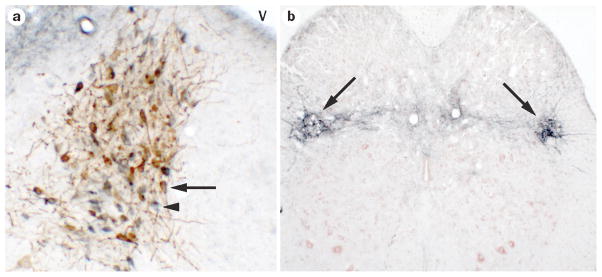
Figure 3.

CRF is a major neurotransmitter in Barrington’s nucleus neurons. A. Brightfield photomicrograph of a rat brain section at the level of Barrington’s nucleus showing CRF-immunoreactive neurons (blue) and neurons that are retrogradely labeled with the tracer fluorogold from the lumbosacral spinal cord (brown). Note that most neurons have the hybrid blue/brown color indicating that they are CRF neurons that project to the spinal cord (example indicated by arrow). The arrowhead points to a neuron that is labeled for CRF only. Dorsal is at the top and medial is to the right. V indicates the fourth ventricle. B. Section at the level of the lumbosacral spinal cord showing dense CRF immunoreactive terminal fields (blue) in the region of the preganglionic parasympathetic neurons (arrows). The section is counterstained with Neutral Red. Dorsal is at the top.
