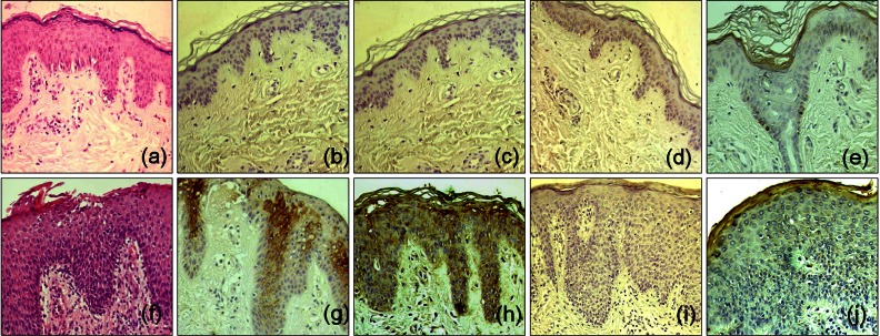
Fig. 1.
Histopathological and immunohistochemical findings of biopsy specimens from normal control (a~e) and atopic dermatitis (AD) patients (f~j). H&E stained sections of the healthy skin of the normal control (a) and skin lesion of an AD patient (f). A variable degree of parakeratosis, acanthosis, spongiosis, and/or exocytosis, and sparse-to-moderate inflammatory cells infiltration were observed (×200). Immunohistochemical reactivity of staphylococcal protein A (SPA), recombinant staphylococcal enterotoxin A (SEA) in the skin of a healthy adult (b, c) and AD patients (g, h). Increased intensity of SPA, SEA immunoreactivity was observed in the upper part of the epidermis from all AD patients in comparison with that of a healthy adult (×200). Immunohistochemical reactivity of recombinant staphylococcal enterotoxin B (SEB) in the skin of a healthy adult (d) and AD patients (I). Minimal immunoreactivity of SEB was observed in the lesional skin of almost all AD patients (×200). Finally immunohistochemical reactivity of toxic shock syndrome toxin-1 (TSST-1) in the skin of a healthy adult (e) and AD patients (j). Mild to moderate immunoreactivity of TSST-1 was detected in the lesional skin of AD patients (TSST-1, ×200).

