
Figure 1.
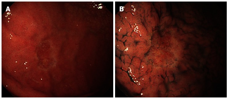
Pre-treatment endoscopic examination. Esophagogastroduodenoscopy revealed pale depressed mucosal lesion on anterior wall of middle gastric body approximately 15 mm in size with no ulcerative finding and non-atrophic background mucosa. A: Conventional white light endoscopy; B: With indigo-carmine dye staining.