Figure 2.

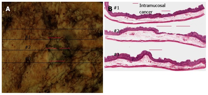
Surgically resected specimen. A: Formalin-fixed specimen from pylorus preserving gastrectomy; B: Panoramic view of lesion with hematoxylin and eosin staining. Mucosal lesion 15 mm × 12 mm in size with no ulcer finding and pink lines corresponding to lesion depression (hematoxylin and eosin staining, × 1).
