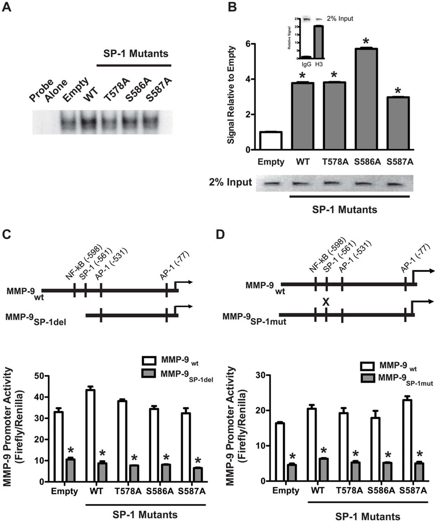
Figure 2. WT and mutant SP-1 bind to SP-1 sites on the MMP-9 promoter.
(A). MH-S cells were transfected with pcDNA3.1.V5.His vectors expressing SP-1WT, SP-T578A, SP-1S586A or SP-1S587A. Control cells were transfected with pcDNA3.1 (empty). Twenty four hours later, nuclei were isolated and subjected to chromatin immunoprecipitation with anti-V5, anti-histone 3 (H3) antibodies or normal rabbit IgG as described in Methods. Immunoprecipitated purified DNA was amplified by real-time PCR using Sybr Green and primers directed against SP-1 binding sites on the MMP-9 promoter or histone binding RPL30 gene. Results show Mean±SEM of signal relative to control and gel separation of PCR products (inset). N=3. (B) MH-S cells were transfected as in A. Nuclear proteins were subjected to EMSA using 32P-labeled consensus SP-1 oligonucleotide as described in Methods. A representative autoradiogram from one out of two representative experiments are shown. (C&D). MH-S cells were transfected with empty, WT or mutant SP-1 vectors together with WT MMP-9 promoter (#2)-, truncated MMP-9 promoter (#4)-(C) or mutated MMP-9 promoter-(D) driven luciferase vectors. Schematic of the MMP-9 promoters is shown (inset). Twenty four hours after transfection, luciferase activity was determined. Results show Mean±SEM of firefly luciferase normalized to Renilla. n=3, *p < 0.05.

