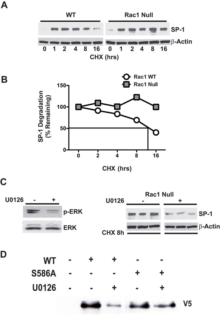
Figure 4. ERK activation increases stability of the SP-1 protein.
(A) WT and Rac1 null macrophages were incubated with 10µM of cycloheximide (CHX) for the indicated times. (B) Densitometric analysis of representative experiments of SP-1 decay in WT and Rac1 null macrophages. (C) Rac1 null cells were incubated for 18 hrs with 0.1% DMSO or U0126 (10µM) and then treated for 8 hrs with CHX (10µM). Immunoblot analysis for p-ERK and SP-1 was performed. (D) Macrophages were transfected with an empty, SP-1WT, or SP-1S586A. Twenty-four hours later cells were cultured in the presence or absence of U0126 for 8 hrs. Immunoblot analysis was performed after affinity purification.

