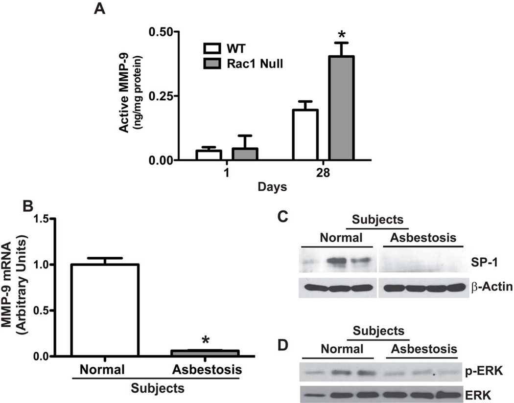
Figure 5. Decreased MMP-9 and SP-1 expression and ERK activation in asbestosis patients.
(A) WT and Rac1 null mice were intratracheally administered 100 µg chrysotile asbestos. Animals were euthanized at one or 28 days after exposure, and BAL was obtained. Active MMP-9 was determined by measuring the difference between total and pro-MMP-9 which were determined. N=4 (WT and Rac1 null); *p < 0.041. (B) Total RNA was isolated from alveolar macrophages obtained from normal subjects (n=3) or patients with asbestosis (n=3). MMP-9 mRNA expression was measured by real-time PCR. * p < 0.001. (C–D) Alveolar macrophages from asbestosis patients (n=3) and normal subjects (n=3) were lysed and subjected to immunoblot analysis for the determination of SP-1 and phosphorylated ERK.

