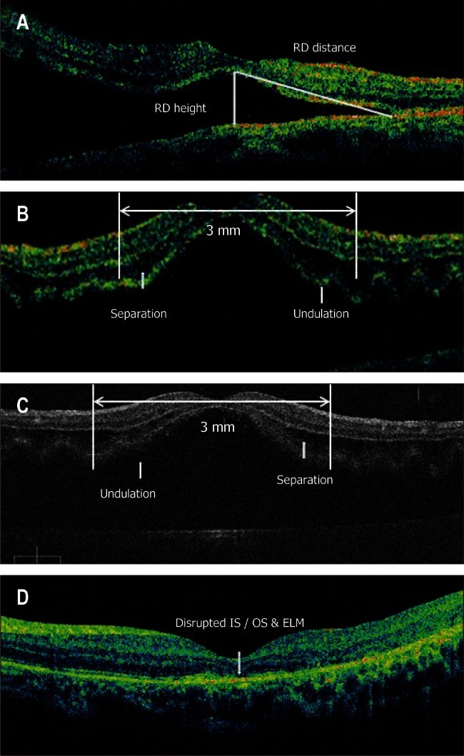
Fig. 1.

(A) time-domain optical coherence tomographic (TD-OCT) image of macula-off rhegmatogenous retinal detachment reveals retinal detachment (RD); the height of such detachment and the extent of RD. (B) A TD-OCT image of macular-off rhegmatogenous retinal detachment shows intraretinal separation (IRS) and outer layer undulation (OLU). (C) An image acquired using spectral-domain optical coherence tomography (SD-OCT). IRS and OLU are relatively large structural changes. Thus, TD-OCT can identify such changes as reliably as can SD-OCT. (D) An SD-OCT image shows disruption of the junction between the photoreceptor inner and outer segments (IS/OS) and an external limiting membrane (ELM); the image was acquired on postoperative follow-up. All parameters were assessed from the central 3-mm diameter area.
