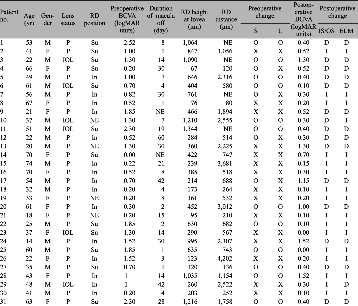
Table 2.
Clinical and optical coherence tomographic characteristics of patients

RD = retinal detachment; BCVA = best-corrected visual acuity; logMAR = logarithm of the minimum angle of resolution; S = intraretinal (outer nuclear layer) separation; U = undulation; IS/OS = junction between the photoreceptor inner and outer segments; ELM = external limiting membrane; M = male; F = female; P = phakic; Su = superior; NE = not evaluable; O = exists; D = disrupted; X = does not exist; I = intact; IOL = intraocular lens (pseudophakia); In = inferior.
