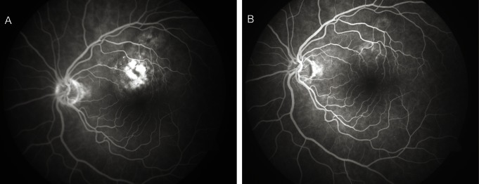
Fig. 2.

(A) Mid-phase fluorescein angiogram of the left fundus showing the relatively well-delineated lesion above the fovea with patchy areas of hyperfluorescence, before transpupillary thermotherapy. (B) Post-treatment early mid-phase fluorescein angiogram demonstrates the almost complete resolution of the lesion.
