Fig. 3.

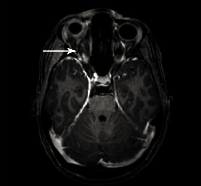
Coronal view on 3-D T1-weighted magnetic resonance imaging after administration of gadolinium reveals the cavernous sinus with thrombosis on both sides, but more pronounced on the right side (arrow).

Coronal view on 3-D T1-weighted magnetic resonance imaging after administration of gadolinium reveals the cavernous sinus with thrombosis on both sides, but more pronounced on the right side (arrow).