
Fig. 4.
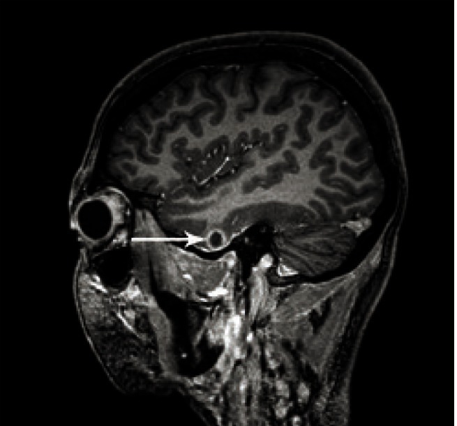
Axial view on a 3-D T1-weighted sequence after administration of gadolinium reveals a thrombosis in the superior ophthalmic vein on the right side (arrow).
Official websites use .gov
A
.gov website belongs to an official
government organization in the United States.
Secure .gov websites use HTTPS
A lock (
) or https:// means you've safely
connected to the .gov website. Share sensitive
information only on official, secure websites.
Axial view on a 3-D T1-weighted sequence after administration of gadolinium reveals a thrombosis in the superior ophthalmic vein on the right side (arrow).