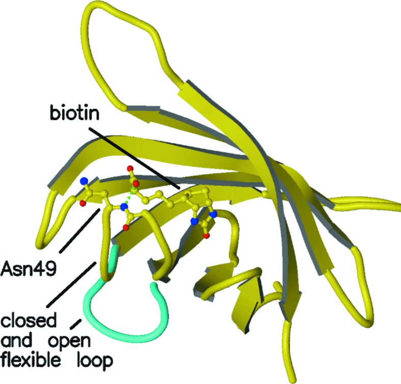
Figure 2.

Overview of the polypeptide trace of a streptavidin subunit with a closed flexible binding loop shown in yellow (subunit A of PDB entry 3ry2). A bound biotin and Asn49 are shown in ball-and-stick mode. The hydrogen bond between the biotin carboxylate and the main-chain amide of Asn49 is shown by a green dotted line, as is the hydrogen bond between Ser45 and the ureido N atom of biotin. The open conformation of the flexible binding loop in unliganded streptavidin is shown in cyan (subunit A of PDB entry 3ry1).
