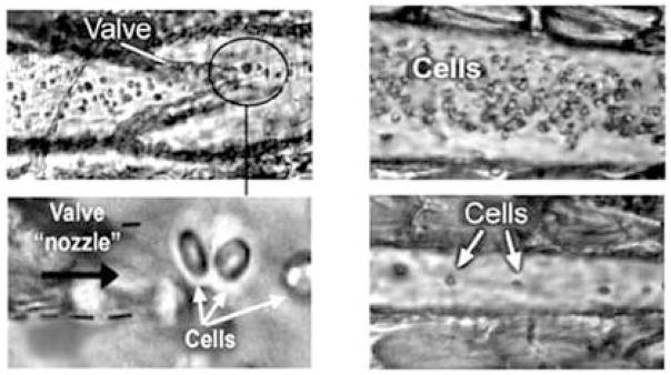
Fig. 2.

High-speed transmittance digital microscopy (TDM) images of cells in the space within the valve area (left) and immediate after a valve (right) of the lymph vessel of rat mesentery at ×10 (top) and ×100 (bottom) magnifications demonstrating the principle of cell flow hydrodynamic focusing. TDM images of cells in the central part of a lymphangion in diastole (top, right) and systole (bottom, right) phases are also shown [from (26)].
