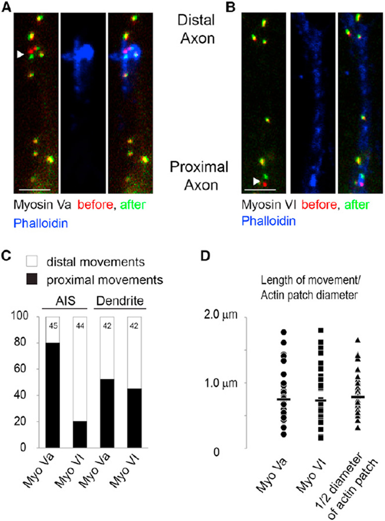
Figure 3. Mapping of Actin Filaments Using Peroxisome Movements Generated by Myosin Motors.
(A) Peroxisomes labeled with GFP and bound to Myosin Va at time point 0 (red), and 60 s later (green). Most peroxisomes did not move and thus are colored in yellow. The arrowhead points to a peroxisome that moved toward the cell body. Note that the distal axon is at the top of the image and the proximal axon is oriented toward the bottom. Phalloidin staining in blue indicates that the peroxisome movement occurred at the same location as an actin patch. Scale bar, 2 µm.
(B) Similar to (A) except that peroxisomes are fastened to Myosin VI. Peroxisome indicated by the arrowhead moves toward the distal axon within an area characterized by high-density phalloidin staining corresponding to an actin patch. Scale bar, 2 µm.
(C) Approximately 80% of peroxisomes in the AIS attached to Myosin Va move toward the cell body, whereas ~80% of AIS peroxisomes attached to Myosin VI move distally. Roughly equal numbers of peroxisomes in dendrites move in both directions when attached to Myosin Va or Myosin VI.
(D) The lengths of peroxisome movements (mean indicated by bar) attached to Myosin Va (circle) or to Myosin VI (square) are comparable to half of the diameter of actin patches measured from SEM images (triangles).
See also Figures S2 and S3.

