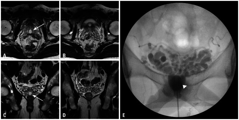
Fig. 2.
T2-weighted magnetic resonance image and seminal vesiculography. (A and C) Approximately 2 cm sized cystic lesion (white arrows) was located in the prostate. (B, C and D) Mild dilatation of the seminal vesicles was observed. (E) The seminal vesicles and midline cyst (white arrow head) were filled with contrast media under seminal vesiculography.

