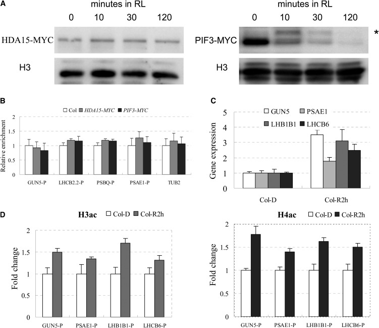
Figure 7.
RL Dissociates HDA15 and PIF3 from Target Genes and Promotes Gene Expression.
(A) Immunoblot analysis of epitope-tagged HDA15 and PIF3 in HDA15-MYC and PIF3-MYC seedlings grown for 2 d in the dark and then maintained in darkness or exposed to RL at 10 µmol m−2 s−1 before extraction. Blotted samples were probed with an anti-myc antibody. Histone H3 was used as a loading control. Asterisk indicates the phosphorylated form of PIF3.
(B) ChIP analysis of the binding of HDA15 and PIF3 to the G-box regions of the promoters in the light-responsive genes when dark-grown seedlings were exposed to RL. P indicates G-box–containing regions in the promoters (as indicated in Figure 5A). The values are shown as means ± sd.
(C) Expression levels of light-responsive genes in Col grown for 2 d in dark and then exposed to RL for 2 h (Col-R2 h) compared with those in dark-grown seedlings (Col-D). The values are shown as means ± sd.
(D) ChIP analysis of histone H3 and H4 acetylation levels of light-responsive genes in Col grown for 2 d in dark and then maintained in darkness or exposed to RL for 2 h. P indicates promoter region. ACTIN2 was used as an internal control. The values are shown as means ± sd.

