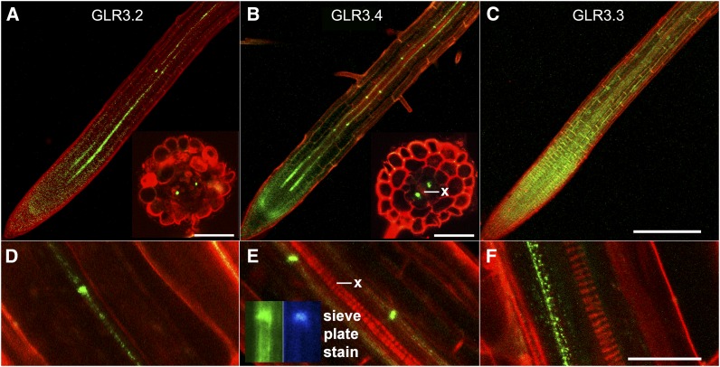
Figure 1.
Localization of GLR3.2, GLR3.3, and GLR3.4 in Roots.
(A) to (C) Confocal microscope images of GFP-tagged GLR3.2, GLR3.4, or GLR3.3 (green) in primary root apices stained with propidium iodide (red) to mark cell boundaries. Insets in (A) and (B) show phloem-localized signal in cross sections cut by hand ∼1 mm from the root tip.
(D) to (F) Higher magnification images of GFP-tagged GLR3.2, GLR3.4, or GLR3.3 signal in mature phloem. Inset in (E) shows that aniline blue, a sieve plate indicator, stains a region of strong GLR3.4-GFP accumulation in the phloem.
Bar in (C) = 200 µm and applies to the main images in (A) to (C); inset bars = 50 µm. Bar in (F) is 20 µm and applies to (D) to (F). x, xylem.

