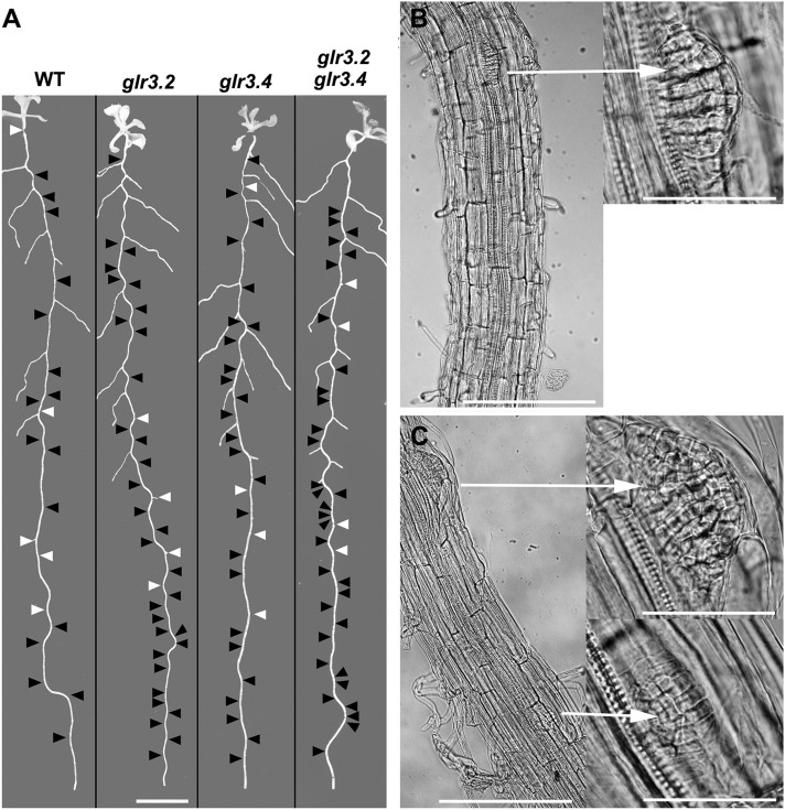
Figure 2.
Spatial Distributions of Lateral Root Primordia and Emerged Lateral Roots in the Wild Type and glr Mutants.
(A) Black arrowheads mark the position of primordia detected by microscopy inspection of the displayed root, and white arrowheads indicate emerged roots not visible in the image. Bar = 8 mm. WT, the wild type.
(B) Segments of primary root showing one lateral root primordium in the wild type and at higher magnification (arrow right).
(C) Two primordia in an equivalent section of a glr3.4 root showing adjacent primordia emerging from the same side of the stele (arrow right).
Bars =200 μm (B) and (C) and 50 μm in the higher magnification images (arrow right).

