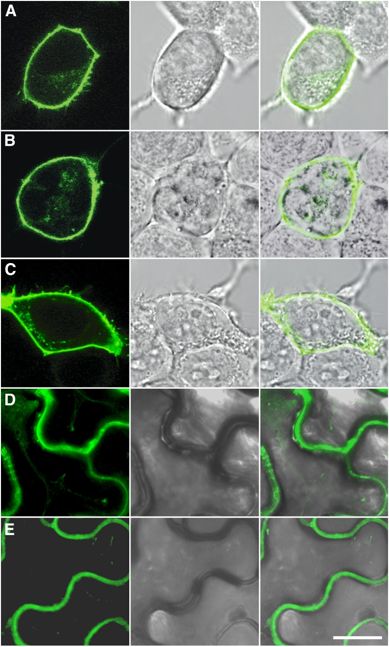
Figure 4.
Subcellular Localization of GLR3.2, GLR3.3, and GLR3.4 in Animal and Plant Cells.
Confocal laser scanning microscope images of HEK293T cells expressing GLR3.2-YFP (A), GLR3.3-YFP (B), and GLR3.4-YFP (C) or N. benthamiana leaf epidermal cells expressing GLR3.2-YFP (D) and GLR3.4-YFP (E) show YFP fluorescence at the plasma membrane. Middle panels show bright-field light micrographs corresponding to the confocal images. Rightmost panels show overlay of the florescence signal with the light micrograph. Bar = 10 μm.

