Figure 1.
Arabidopsis TEN1 Is a Member of CST Complex.
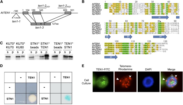
(A) Schematic of TEN1 gene structure. The T-DNA insertion in ten1-1 is illustrated, along with the position of two antisense constructs and the point mutation responsible for the G77E mutation in ten1-3. aa, amino acids.
(B) Alignment of TEN1 proteins from different eukaryotes. At, Arabidopsis thaliana; Os, Oryza sativa (rice); Pt, Populus trichocarpa (poplar); Mm, Mus musculus; Hs, Homo sapiens; Sp, Schizosaccharomyces pombe. The positions of β-strands of the OB-fold are indicated below the alignment. Green, 100% similarity; chartreuse, 80 to 99% similarity; yellow, 60 to 79% similarity; and gray, below 60% similarity.
(C) TEN1 interacts with STN1 in vitro. Results of coimmunoprecipitation performed with recombinant proteins. One protein is [35S]Met labeled (asterisk), and the other is T7 tagged and unlabeled. s, supernatant; p, pellet. Results for the positive (KU70/KU80) and negative (KU70/KU70) controls are shown.
(D) Yeast two-hybrid assay results for STN1 and TEN1. The two proteins fused to GAL4-AD and GAL4-BD were coexpressed and grown on selection plates for His auxotrophy (left) or assayed to detect β-galactosidase activity of positive transformants (right). “−” Indicates empty vector.
(E) Nuclear localization of TEN1 in purified nuclei. TEN1 was detected by anti-TEN1 antibody in hexaploid Arabidopsis suspension cell culture. Telomeres were labeled by FISH using a rhodamine-labeled telomere probe. 4′,6-Diamidino-2-phenylindole (DAPI)–stained nuclei are shown. In the merge, closed white arrows denote subcentromeric stretches of telomeric DNA on chromosome 1. TEN1 colocalization with telomeres is indicated by the open white arrow.

