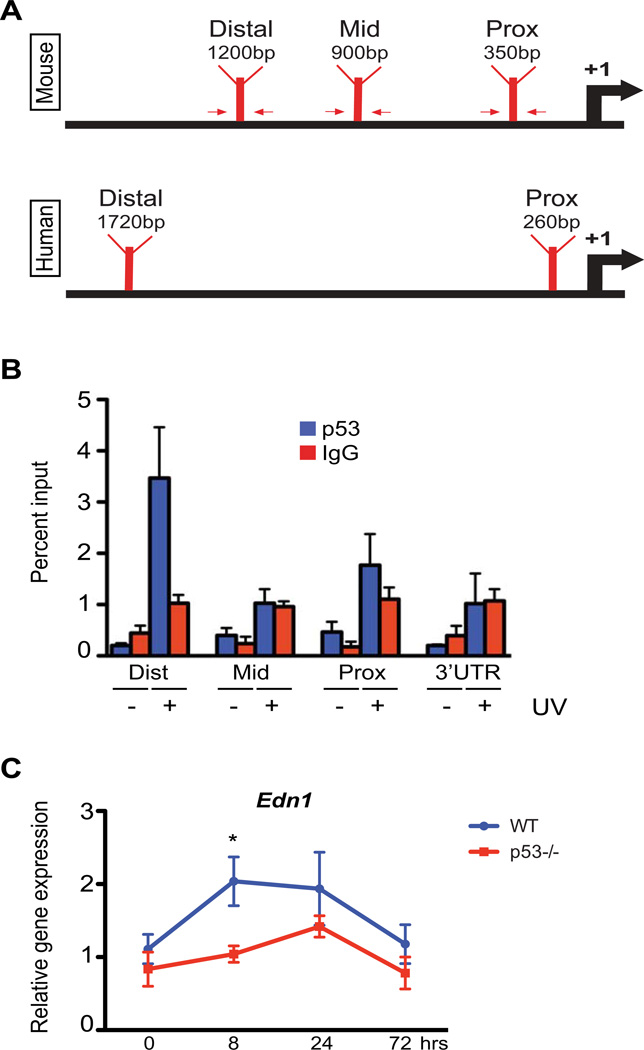
Figure 1.
Positive regulation of Edn1 expression after UV exposure by p53 in murine keratinocytes. (A) Schematic of predicted p53 binding locations on murine and human Edn1/EDN1 promoters. Arrows indicate primers designed for chromatin immunoprecipitation (ChIP). (B) ChIP assay on primary murine keratinocytes using anti-p53 antibody following presence or absence of UV exposure. Results were analyzed by qPCR using primers specific to proximal, mid and distal regions (indicated in A). Primers directed against the 3’ UTR region of EDN1 and non-specific IgG antibody were used as negative controls. (C) Relative gene expression of Edn1 in epidermis from adult wildtype C57BL/6 and p53−/− mice at designated time points post-UV exposure. All experiments were done using a minimum of three biological replicates from each group and in all cases are expressed as mean +/− SEM. Statistical analysis was performed using Graphpad Prism, * = p < 0.05.

