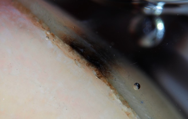
Figure 2.

Dermatoscopy of the free edge of the nail plate shows that melanin has been preferentially incorporated into the deeper portion of the nail plate. This indicates that it is originating from a lesion in the distal portion of the nail matrix.
