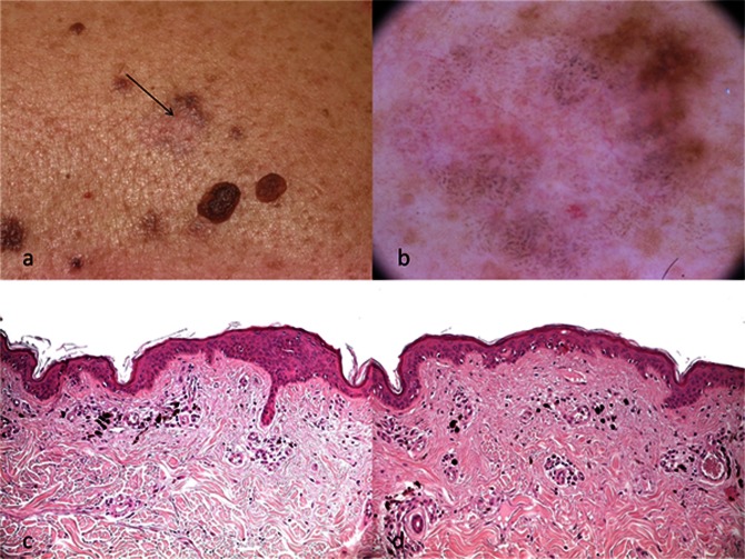
Figure 2.
(A) Clinical image showing a fully regressed melanocytic nevus (arrow). (B) Dermoscopy reveals diffuse blue-gray granules, white areas and some telangiectatic vessels. Remnants of pigmented network can be observed at the upper right part of the lesion. (C, D) Scattered melanophages, prominent fibrosis and telangiectasias can be seen on histopathology, corresponding to the above-mentioned dermoscopic features (H&E ×20 magnification). [Copyright: ©2012 Lallas et al.]

