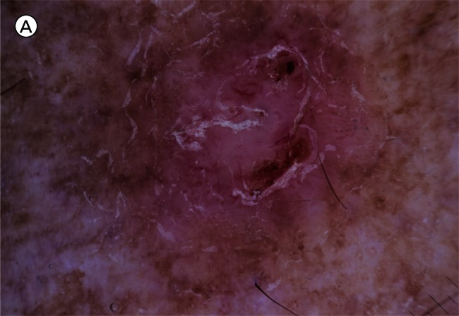
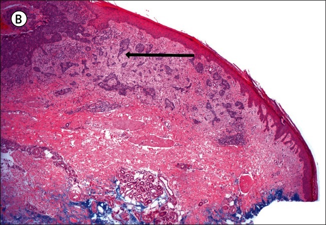
Figure 4.
A) Infiltrating basal cell carcinoma: dermatoscopy. [Copyright: ©2012 Pyne et al.] B) Infiltrating basal cell carcinoma: histopathology (same lesion as Figure 4A). Hematoxylin and eosin stain. Black arrow to the collagen rich tumor stroma. [Copyright: ©2012 Pyne et al.]


