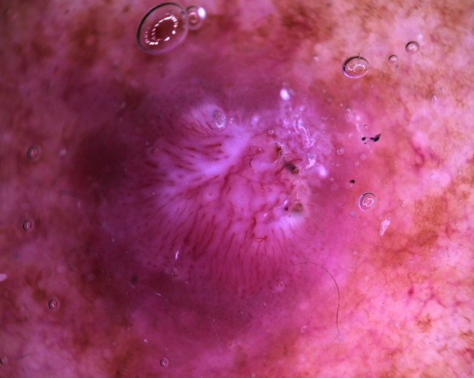
Figure 4.

Dermatoscopy of a well differentiated SCC on the shoulder, orderly peripheral dot vessels merging into more central hairpin or loop vessels. The central tumor area has an elevated surface with white structureless areas. Vessels displaying a while halo are easily identified. [Copyright: ©2012 Pyne et al.]
