
Figure 6.
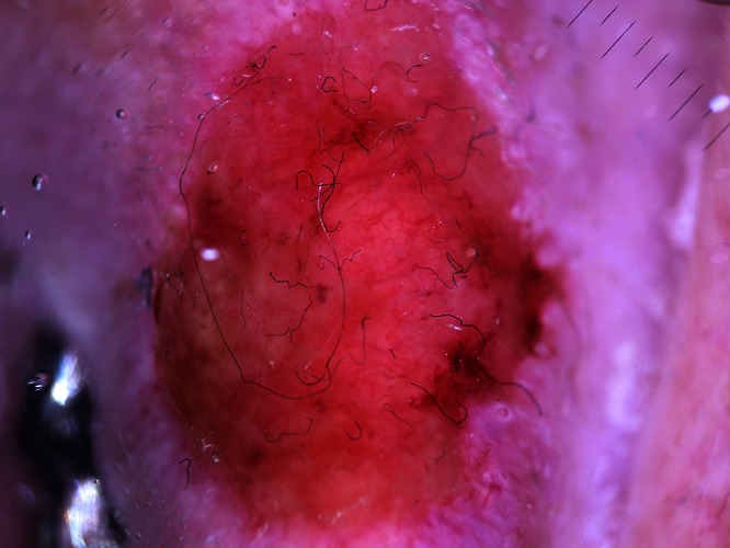
Dermatoscopy of a poorly differentiated SCC on the ear; the tumor surface follows the cartilage contours; pink areas cover the whole tumor surface; branching vessels without a white halo are common. [Copyright: ©2012 Pyne et al.]
Official websites use .gov
A
.gov website belongs to an official
government organization in the United States.
Secure .gov websites use HTTPS
A lock (
) or https:// means you've safely
connected to the .gov website. Share sensitive
information only on official, secure websites.
Dermatoscopy of a poorly differentiated SCC on the ear; the tumor surface follows the cartilage contours; pink areas cover the whole tumor surface; branching vessels without a white halo are common. [Copyright: ©2012 Pyne et al.]