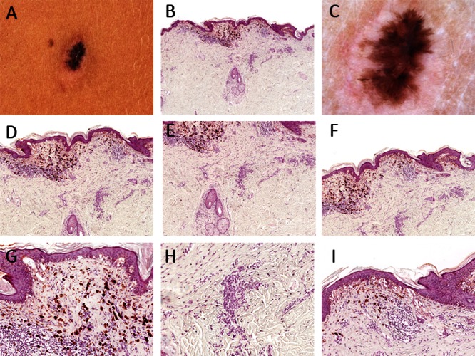
Figure 3.

(A & C) Clinical and dermatoscopic picture of a brown papule on the back. Dermatoscopically one can see segmental radial lines within a hypopigmented structureless area (scar). (B & D–H) Dermatopathologic images of the lesion shown in A & C. Melanocytes in the epidermis are arranged as single cells and in confluent nests. Inconspicuous nests of small melanocytes arranged in an adnexocentric fashion can be spotted in the dermis. [Copyright: ©2013 Tschandl.]
