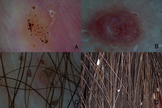
Figure 2.
Entomodermoscopy improves the diagnosis of skin infestations and infections. (A) Stereotypical appearance of plantar viral wart showing structureless white-yellow areas and multiple small linear brown to red dots and streaks (=splinter hemorrhages). (B) Molluscum contagiosum is typified by central opaque yellowish globules, which are surrounded by blurred linear vessels (=crown vessels). (C) Dermoscopy of phthiriasis pubis with vital nits attached to the hair shaft. Vital nits have a convex end and appear translucent brownish. (D) Dermoscopy of pseudo-nits due to hair casts, which appear as white amorphous structures attached to the hair shaft. Original magnification x10. [Copyright: ©2013 Zalaudek et al.]

