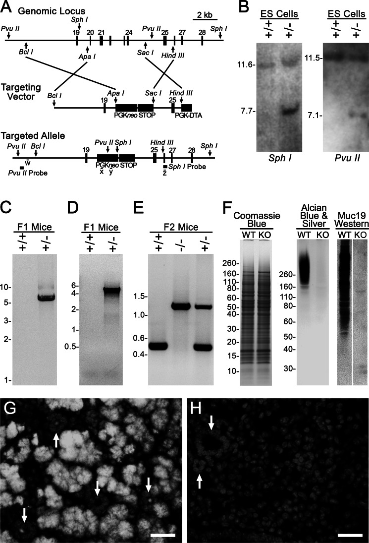
FIGURE 2.
Production and characterization of Muc19 knock-out mice. A, strategy for targeted disruption of Muc19 transcripts from the gene Muc19/Smgc. The homology arms were inserted into the selection cassette PGKneolox2DTA that encodes for neomycin under control of the PGK promoter. The PGKneo sequence is flanked by loxP sites (not shown). The sequence from the STOP cassette, pBS302, was inserted between the downstream loxP site, and diphtheria toxin A was driven by the PGK promoter for negative selection. The targeted allele is shown with restriction sites (PvuII and SphI) and probes used for Southern blot analyses as well as sites of PCR primers w–z. B, Southern blot analyses of DNA from nonrecombinant ES cells (+/+) and correctly targeted ES cells (+/−) digested with SphI and with PvuII. C, PCR-based genotyping of F1 agouti mice to distinguish the absence (+/+) or presence (+/−) of germ line transmission and correct insertion of the 3′-end of the targeted allele using primers w and x (as shown in A). D, same as in C, but using primers y and z to detect germ line transmission and correct insertion of the 5′-end of the targeted allele. E, genotyping of progeny from intercrossing F1 recombinant mice. F, sublingual gland homogenates (wet weight, 200 μg) from wild type (WT) and homozygous Muc19 knock-out (KO) mice were subjected to SDS-PAGE (4–12% gel) and stained with Coomassie Blue to detect proteins. Homogenates (wet weight, 300 μg) were also run on a 4–12% gel and either stained with Alcian blue and subsequent silver enhancement to detect highly glycosylated glycoproteins or identical lanes blotted and probed with rabbit anti-Muc19. G and H, anti-Muc19 immunofluorescence. G, low power micrograph of a paraffin section from a 3-day-old wild type mouse probed with rabbit anti-Muc19 and Alexa Fluor 568 goat anti-rabbit IgG displaying Muc19 in mucous cells of tubuloacini. The nuclei were counterstained with DAPI (light gray). Arrows indicate cross-sectioned ducts, most of which contain Muc19 within their lumen. H, section from a 3-day-old Muc19 KO mouse processed in an identical manner displays an absence of Muc19 immunostaining. Only DAPI staining is visible. The arrows indicate cross-sectioned ducts. Bars, 25 μm.

